- SMC Laboratories, Inc. >
- Investment in Biotechnology Business
Business Description
01
Investment in Biotechnology Business
SMC Laboratories, Inc. is a Tokyo-based nonclinical Organization (CRO) that specializes in research on fibrosis and inflammation. We have more than 700 clients which include pharmaceutical companies, universities, and national medical institutions, and have assisted with nonclinical trial applications for more than 10 pipeline drugs, mainly for nonalcoholic steatohepatitis (NASH).
SMC Laboratories, Inc.
Head Office: Technoport Clinic Kamata, 2-16-1 Minami-Kamata, Ota City, Tokyo, Japan
https://www.smclab.co.jp/


SMC Laboratories’ Contracted Business Focused on Nonclinical CRO Portfolio
NAFLD / NASH
・NASH-HCC: STAM™ Model
・Fatty Liver / NAFLD: MCD Model
・Fatty Liver / NAFLD: ob / ob Model
Inflammation and Fibrosis
IPF: Bleomycin-induced lung fibrosis model
COPD: Porcine Pancreatic Elastase (PPE) model
Liver cirrhosis: CCl4-induced liver fibrosis model
Cholestatic liver disease: Bile duct ligation (BDL) model
CKD: UUO-induced kidney fibrosis model
Adriamycin-induced nephropathy model
IBD: DSS-induced colitis model
Alzheimer’s disease: icv-STV model
Systemic sclerosis: Bleomycin-induced skin fibrosis model
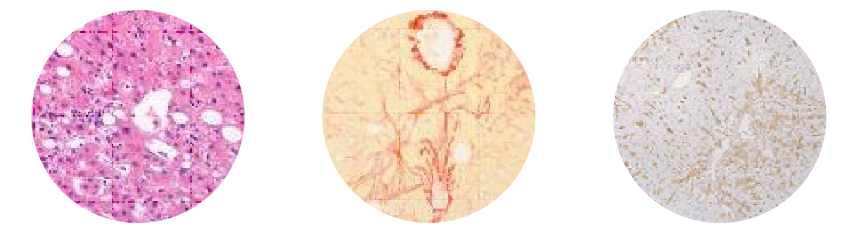
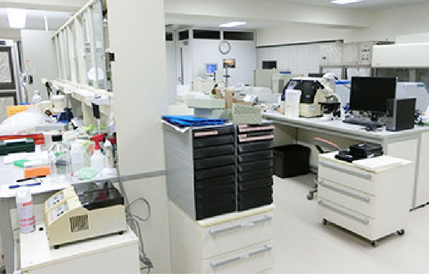
Histopathology and Imaging Services
Processing and Embedding
・Embedding of human and animal tissues in paraffin (FFPE blocks) from fixed tissues.
・Embedding of human and animal tissues in O.C.T. (frozen O.C.T. embedded tissues) from fixed tissues.
・Instruction in preparing fresh frozen blocks for frozen sections.
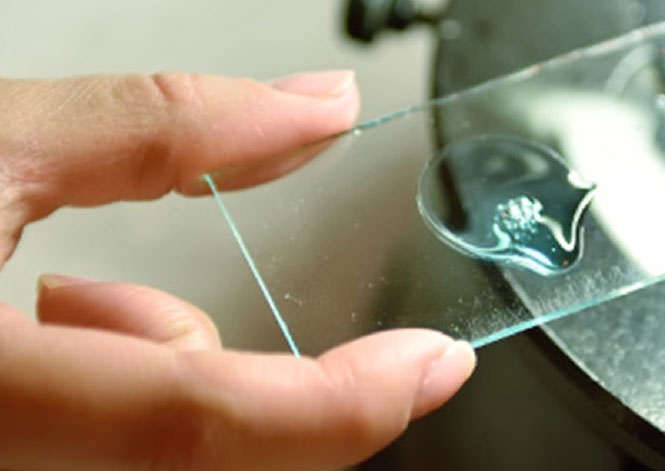
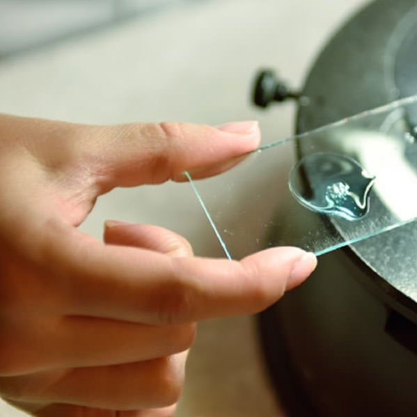
Sectioning
・Cutting of 4-8 μm thick sections from paraffin- and OCT-embedded blocks.
・Preparation of serial thin sections. Routine Staining.
・HE, Sirius Red, Masson’s trichrome, PAS, Oil Red, etc.
Reporting
We can perform evaluations in a variety of disease models.
Immunohistochemistry (IHC)
Inflammation-related molecules, fibrosis-related molecules, etc.
・Functional Immunohistochemistry (double / multiple staining).
・Optimization and validation of new antibody staining protocols.
Image Analysis
・Area, length, diameter, stained cand ell count, percentage of positive area, and shape, etc.
・Proliferation, apoptosis, pathological grading, etc.
Results Interpretation
Discussion of histological findings from a pharmacological perspective.